

Faculty Advisor(s)
Elaine Halesey
Files
Abstract
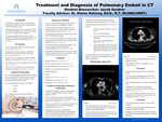
This research project elaborates on the diagnosis and treatment of pulmonary emboli, and how computed tomography can provide a quick and accurate diagnosis. Statistics, symptoms, computed tomography pulmonary angiography protocol, and pulmonary emboli radiographic appearance are also discussed to provide information on how a diagnosis is achieved and which treatment is used. Pulmonary emboli are a life-threatening condition that, if untreated, can potentially be fatal. Pulmonary emboli are the third most frequently occurring cause of cardiovascular death behind a stroke and myocardial infarction. Deaths in the United States from pulmonary emboli are approximately 300,000 deaths per year. In addition, 5-10% of in-hospital deaths are caused by pulmonary emboli. Computed tomography is the quickest and most effective method for diagnosing pulmonary emboli. Specifically, computed tomography pulmonary angiography is used which highlights the pulmonary artery using iodinated contrast media. Treatments vary according to severity. In most cases, patients are given anticoagulation medication which is used to prevent blood clots. In more severe cases, patient will require surgery to remove the emboli called an embolectomy. Further research should be conducted for finding new treatment methods to lower the death toll in the United States.
Publication Date
2024
Document Type
Poster
Department
Medical Imaging
Keywords
pulmonary embolism, computed tomography, computed tomography pulmonary angiography, pulmonary embolism treatment, pulmonary embolism diagnosis
Disciplines
Medicine and Health Sciences | Radiology
Recommended Citation
Gardner, Jacob, "Treatment and Diagnosis of Pulmonary Emboli in CT" (2024). Student Research Poster Presentations 2024. 1.
https://digitalcommons.misericordia.edu/research_posters2024/1