

Faculty Advisor(s)
Lynn Blazaskie
Files
Abstract
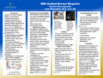
Magnetic resonance imaging (MRI) is a non-invasive technique that produces computer-generated cross-sectional images. The images produced contain anatomic and physiologic information. Using non-ionizing radiation, MRI creates images through the interaction of magnetic fields and radio frequency energy on biologic tissue. The specificity present in the images produced is the main reason MRI is a popular choice of diagnostic testing. MRI is used to evaluate organs, tissues, and skeletal systems and different sequences are used depending on what the main focus of the test is. MRI images are more complex because they contain information about different properties of tissue-proton density, relaxation rates, and flow phenomena. MRI breast imaging is performed on women who are at high risk of developing breast cancer, or dense breasts because it is a highly detailed test. The different sequences used could detect small abnormalities that may be missed under ultrasound or mammography. MRI guided breast biopsies are done when an abnormality is detected and a preoperative step in treating the disease. The specialized equipment is helpful in determining the exact size, location, and extent of the cancer before choosing a path of treatment. MRI guided breast biopsies are becoming more prevalent in diagnosing and planning treatment for women who have been diagnosed with breast cancer because of the specificity.
Keywords: Magnetic resonance imaging, non-ionizing radiation, MRI breast imaging, MRI guided breast biopsy
Publication Date
2022
Document Type
Poster
Department
Medical Imaging
Keywords
magnetic resonance imaging, non-ionizing radiation, MRI breast imaging, MRI guided breast biopsy
Disciplines
Medical Sciences | Medicine and Health Sciences
Recommended Citation
Roccograndi, Rachel, "MRI Guided Breast Biopsy" (2022). Medical Imaging Senior Posters. 37.
https://digitalcommons.misericordia.edu/medimg_seniorposters/37