

Faculty Advisor(s)
Anthony Serino
Files
Download Full Text (24.1 MB)
Abstract
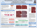
The liver is an organ with many functions in the body, including storage of essential nutrients, detoxification of blood, and digestion and metabolism. The major functional unit of the liver is the liver lobule, which contains the micro-anatomy of the liver, including hepatocytes, sinusoids, portal triads, and the central vein. The liver is a resilient organ that has the capability to regenerate up to two-thirds of its tissue. In cirrhosis, a condition in which collagen is produced and results in fibrotic tissue and causes normally functioning tissue to become scar tissue, the liver is no longer capable of its regenerative functions. By taking a biopsy from the patient’s liver, there are several cellular histopathological indicators of cirrhosis that can confirm the stage of cirrhosis the patient is in. Some of the indicators include the increased presence of Kuppfer cells and hepatic stellate cells, the decreased presence of liver sinusoidal epithelial cells, and the formation of tissue nodules and fibrotic bands. Histological microscope slides were created and imaged to determine if the histopathological indicators for cirrhosis were present and then correlated with the size of fat pads in the cadavers the samples were obtained from. It was determined there is a slight positive correlation between estimated percentage of cirrhosis and size of pubic fat pads.
Publication Date
2023
Document Type
Poster
Department
Biology
Keywords
liver cirrhosis, liver cirrhosis in cadavers, histopathological indicators of cirrhosis, histopathological markers of cirrhosis, correlation study, cadaver fat pad size
Disciplines
Biology | Life Sciences
Recommended Citation
Wrobleski, Victoria, "Histopathological Markers of Liver Cirrhosis Correlating with Fat Pads in Cadavers" (2023). Student Research Poster Presentations 2023. 24.
https://digitalcommons.misericordia.edu/research_posters2023/24