

Faculty Advisor(s)
Elaine Halesey
Files
Abstract
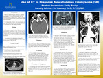
Subcutaneous emphysema is a follow up symptom to a traumatic event, surgery, or nose-blowing. In addition subcutaneous emphysema is considered benign and self limiting. Computed tomography (CT) is the imaging modality of choice for diagnosing subcutaneous emphysema. CT is preferred because of its sensitivity to tissue densities. A CT will demonstrate dark air pockets within the subcutaneous layer of the skin. Furthermore, upon palpation of the the skin a crepitus crackling sound is demonstrated. Moreover, listening to the skin with a stethoscope will emit a high-frequency acoustic sound. Nevertheless, extensive subcutaneous emphysema may require medical assistance. Typically, orbital subcutaneous emphysema is non-life threatening and will resolve within a few days as the body absorbs the air. If the air is extensive and compresses on the optic nerve medical attention is required to prevent vision loss. In addition, traumatic subcutaneous emphysema may be caused from pneumothorax or other blunt trauma. The air rises and spreads within the body and can travel though different anatomical planes. Lastly, subcutaneous emphysema may be a complication of surgical procedures. Individuals are susceptible to suffering from subcutaneous emphysema after procedures like colonoscopies. To conclude, subcutaneous emphysema is a symptom that can be diagnosed by CT and is relatively harmless.
Keywords: Subcutaneous emphysema, CT, benign, crepitus
Publication Date
2022
Document Type
Poster
Department
Medical Imaging
Keywords
subcutaneous emphysema, CT, benign, crepitus
Disciplines
Medicine and Health Sciences
Recommended Citation
Paciga, Emily, "Use of CT to Diagnose Subcutaneous Emphysema (SE)" (2022). Medical Imaging Senior Posters. 26.
https://digitalcommons.misericordia.edu/medimg_seniorposters/26