

Faculty Advisor(s)
Elaine Halesey
Files
Abstract
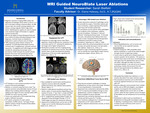
This project explains how magnetic resonance imaging (MRI) is used for neuroblate laser ablations, or laser interstitial thermal therapy (LITT). LITT therapy involves the usage of heat to treat intracranial pathologies such as glioblastomas. Laser therapy is an non-invasive alternative to open resection and craniotomy procedures in which a surgical opening of the skull and brain tissue are necessary. Ablations are considered a safer alternative because patients showed no signs of declining cognitive functions post surgery. Magnetic resonance scanners are used in conjunction with laser systems. The two types of systems are Visualase (Visualase, INC.) and NeuroBlate (Monteris Medical, Inc.) that demonstrates the temperature of surrounding tissue in both the central and peripheral zones of the brain. Patient is under general anesthesia while a “burr” hole is drilled into their head for entry of the laser probes. MRI uses T1 sequences to demonstrate the full length of the probe and FLAIR sequences are utilized for anatomical reference. The ablated lesion can be seen in the axial view of a MR image; then diminishes in size up to three months.
Keywords: Magnetic Resonance Imaging (MRI), Neuroblate Laser Ablation, Laser Interstitial Therapy (LITT), Visualase, NeuroBlate, Glioblastoma
Publication Date
2022
Document Type
Poster
Department
Medical Imaging
Keywords
magnetic resonance imaging, NeuroBlate, ablations
Disciplines
Medicine and Health Sciences
Recommended Citation
Bielfeld, Sarah, "MRI Guided NeuroBlate Laser Ablations" (2022). Medical Imaging Senior Posters. 30.
https://digitalcommons.misericordia.edu/medimg_seniorposters/30