

Faculty Advisor(s)
Elaine Halesey
Files
Abstract
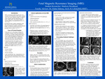
This project explains fetal magnetic resonance imaging (MRI) and its role in prenatal diagnosis. It discusses various aspects, including types of prenatal imaging, clinical applications, the imaging procedures, safety considerations, and technical challenges. Understanding the benefits and limitations of fetal MRI allows for earlier and more accurate detection of fetal abnormalities. Fetal MRI serves as a secondary imaging modality that complements ultrasound by providing detailed imaging of fetal anatomy. Several congenital anomalies including those involving the central nervous system (CNS), as well as the abdominal and thoracic regions, can be thoroughly evaluated to provide additional information and confirm diagnoses made using ultrasound. Safety concerns such as tissue heating, acoustic damage, and contrast agent use are evaluated toensure no harm to the fetus. Advances in artificial intelligence (AI) and the use of 3 T scanners over 1.5 T have significantly improved image resolution and reduced motion artifacts. Overall, fetal MRI is improving prenatal diagnosis through ongoing technological advancements.
Publication Date
2025
Document Type
Poster
Department
Medical Imaging
Keywords
fetal magnetic resonance imaging, prenatal diagnosis, ultrasound, artificial intelligence (AI), image resolution, motion artifact
Disciplines
Medicine and Health Sciences
Recommended Citation
Woodhead, Madison, "Fetal Magnetic Resonance Imaging (MRI)" (2025). Medical Imaging Senior Posters. 59.
https://digitalcommons.misericordia.edu/medimg_seniorposters/59