

Student Research Poster Presentations showcase innovative research by Misericordia students across all disciplines.
-

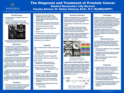
The Diagnosis and Treatment of Prostate Cancer
Lilly Bernard
The purpose of this project is to discuss all aspects of prostate cancer, with a focus on radiation therapy as a treatment option. An overview of the disease is provided, including statistics, risk factors, symptoms, the process of diagnosis, treatment options, and a case study. Prostate cancer is the most common cancer in men and second leading cause of cancer death in the United States, making it widespread and deadly. This means that it is important to understand as much as possible about the disease in order to achieve the best outcomes possible. The project lists experiences from patients who have undergone radiation therapy, touching on their experiences during and after treatment. Stereotactic body radiation (SBRT) is a recent development in radiation therapy that allows for a high dose to be delivered to a small area. The case study follows a patient who has recurrent prostate adenocarcinoma and receives SBRT as treatment.
-

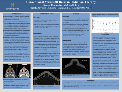
Conventional Versus 3D Bolus in Radiation Therapy
Madison Elizabeth Brdaric
Radiation therapy is a form of cancer treatment that uses high-energy radiation to target tumors while protecting healthy tissue. Tumors near the skin can be difficult to treat because the surface does not get enough radiation. Boluses are tissue-equivalent materials that help focus radiation on the tumor. Conventional boluses work well on flat surfaces but struggle on awkward or uneven surfaces such as the nose or ear, resulting in air gaps that limit the efficiency of the treatment. 3D printed boluses solve this problem because they are custom-made to fit each patient's unique body structure. This allows the appropriate amount of radiation to be delivered exactly where it is required. 3D printed boluses also save time during treatment setup while improving patient outcomes. While 3D printed boluses take longer to manufacture and require additional long-term research, they are a promising tool for treating cancers near the skin. The type of bolus used should be determined on a patient-by-patient basis, as each patient's anatomy and tumor location vary.
-

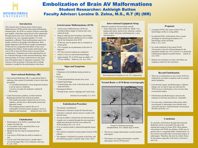
Embolization of Brain AV Malformations
Ashleigh M. Button
Abstract
The purpose of this research is to discuss brain Arteriovenous Malformations (AVM) and the Interventional Radiology treatment plan. An AVM is a section of blood vessels that gets tangled, which then causes blood to flow abnormally through arteries and veins in the brain. Brain AVMs are considered to be very rare and approximately 1% of the population is affected. These malformations seem to be equally common in all genders and ethnicities. Most people diagnosed with AVMs are usually asymptomatic. AVMs may be a congenital birth defect or may occur throughout the lifetime. Endovascular embolization uses coils to fill or close the blood vessel to prevent future complications (Cleveland Clinic, 2022). Treatment such as this embolization can be conducted through an Interventional Radiology short term procedure involving a small incision in the groin area is available if the patient starts to experience symptoms. The outcome of this procedure is the hope that no other further treatment is needed for the embolization in the brain.
-
 Protocols for Older Adults with Cardiovascular Diseases: A Systematic Review by Matthew G. Carter, Emma Pole, Elizabeth Buonanno, Abigail Curtis, and Nicole Paranich")
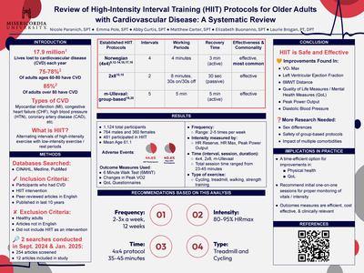
Review of High-Intensity Interval Training (HIIT) Protocols for Older Adults with Cardiovascular Diseases: A Systematic Review
Matthew G. Carter, Emma Pole, Elizabeth Buonanno, Abigail Curtis, and Nicole Paranich
Background: Cardiovascular disease (CVD) is the leading cause of death worldwide, with approximately 17.9 million lives lost each year, and has an incidence of 77-80% amongst those 60-80 years old. HIIT is not the initial treatment of choice in those with CVD among physical therapists because of the fear and concern relating to the patient’s response to high-intensity exercise.
Objective: The purpose of this systematic review was to determine the effectiveness of high-intensity interval training (HIIT) on older adults with cardiovascular disease and, if it was effective, find the proper protocol for this population. This systematic review intends to find the proper frequency, intensity, time, and type (FIIT) of HIIT for these patients to receive the maximal benefits in the safest way possible.
METHODS: The search for this systematic review took place from September of 2024 to January of 2025 and used CINAHL, PubMed, and Medline databases. The search terms within these databases included: high intensity interval training, HIIT, older adults, protocol, cardiovascular, elderly, and heart disease. The inclusion criteria of these searches included participants with cardiovascular diseases, HIIT interventions, peer-reviewed, randomized controlled trials, English language, and published within the last 10 years. After a thorough review of the mentioned databases using these criteria, 12 studies were chosen to be included in this systematic review.
RESULTS: The results of the chosen articles showed that HIIT was more beneficial for older adults with cardiovascular disease compared to interventions of moderate-continuous training (MCT) and control groups with no adjustments to their current exercise regimen. HIIT showed greater improvements in older adults' peak VO2 max and left ventricular ejection fraction (LVEF), as well as increases in 6 MWT and QOL scales. Of the HIIT protocols examined, the most beneficial protocols were the 4x4, consisting of 4 minute high-intensity training, followed by 3 minutes of active/passive rest for 4 sets total, as well as the 2x8, using 8 minutes of high intensity training with 30 seconds of high intensity training, followed by 30 seconds of active rest, for 2 sets. There were also some benefits seen with the M-ullevaal protocol, which is 3 HIIT sets at 90% maxHR, combined with 2 moderate-continuous training (MCT) sets at 70% max HR. These protocols were performed through various activities, including walking, cycling, elliptical, and muscle strengthening exercises.
CONCLUSION: There were significant physiological and psychological benefits seen amongst the older adults participating in HIIT training. Throughout a review of the 12 studies, there was little to no increase in adverse events amongst the HIIT group compared to the MCT or control groups, helping to emphasize the fact that these protocols can be performed in a safe and effective manner for this patient population. There was also an overall beneficial frequency, intensity, time, and type that can be used as a baseline for future intervention within this population. In the future, studies can be done to evaluate the difference between males and females with HIIT training, as well as studies to determine the proper intensity thresholds to reach HIIT.
-

Physical and Perceived Effects of the Menstrual Cycle on Female Athletes
Talia E. Casarella, Alyssa M. Quiteles, Emily E. Liddick, Madelyn R. Swarthout, and Rebecca J. Turner
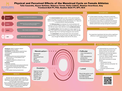
Purpose: The purpose of this study was to understand the physical and perceived effects of the menstrual cycle on female athletes’ performance during training and competition.
Hypothesis: The researchers hypothesized that there would be a significant difference in performance based on the phase of the menstrual cycle.
Methods: This study used EBSCO Academic Search Complete and Science Direct, focusing on peer-reviewed articles from the last 10 years. Search terms included: (exercise or physical fitness or physical activity) AND (menstrual cycle or period or menstruation or menses or follicular phase or luteal phase) AND (female athletes or women athletes or athletic females or sportswomen) AND (competitive or competition). Participants were female athletes experiencing a menstrual cycle, with or without hormonal contraception. Inclusion criteria required studies to assess performance, physical function, and/or mental status across menstrual cycle phases and include competitive athletes, with a potential control group of non-competitive athletes. Exclusion criteria eliminated non-peer-reviewed articles, studies involving male subjects, or those not assessing relevant functions or focusing on menstrual dysfunction. Study quality was assessed using the Hierarchy of Evidence Scale.
Results: In 2024, a comprehensive search using EBSCO and ScienceDirect databases yielded 441 articles, with 9 selected for inclusion and 3 additional articles identified through hand searches. Most studies were classified as level 3 on the hierarchy of evidence, while 2 were deemed not applicable due to qualitative design. In 2025, a similar search produced 83 articles, resulting in 4 included studies and 1 from a hand search. Among these, one was a level 2 experimental study, one was not applicable, and one was a qualitative descriptive study, which was included to provide further insight.
Conclusion: This systematic review highlights the influence of the menstrual cycle on female athletes' performance, with effects varying across different phases. During the follicular phase, women reported lower serenity levels compared to men but exhibited greater motivation during menstruation than their male counterparts. The luteal phase was marked by mood declines in approximately 60% of women, leading to reduced motivation and performance, with these effects often persisting into early menstruation. Additionally, studies found that lactate concentration was higher in the late follicular phase, while endurance capacity declined during the luteal phase. These findings suggest that different menstrual phases influence energy utilization, potentially affecting fatigue levels and recovery rates in female athletes. In conclusion, this review underscores the need for further research to deepen our understanding of menstrual cycle-related performance fluctuations and highlights the importance of integrating these considerations into training and rehabilitation programs.
Clinical Relevance: This study's clinical relevance for physical therapists lies in its potential to guide evidence-based care for female athletes. Understanding how menstrual cycle phases impact performance and physical function can help tailor rehabilitation and training programs. This research can also inform strategies to manage menstrual-related changes, optimizing care, injury prevention, and performance outcomes for female athletes.
-

The Effects of Vestibular Rehabilitation Interventions on Patients with Parkinson's Disease: A Systematic Review
Christopher Farrell and Eduard Turner
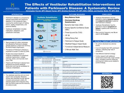
Background: Vestibular rehabilitation is known to be effective in the treatment of vestibular dysfunction. While patients with Parkinson's disease have both central and peripheral vestibular dysfunction, there is little information regarding the efficacy of vestibular rehabilitation for the treatment of these individuals.
Study Design: Systematic Review
Purpose: The purpose of this systematic review was to examine the effects of vestibular rehabilitation in patients with Parkinson’s disease.
Results: The interventions identified in this study resulted in improved Berg Balance Scale (BBS) and Dizziness Handicap Questionnaire (DHI) scores.
Conclusion: Vestibular rehabilitation improves balance and subjective dizziness in patients with Parkinson’s disease.
-

The Role and Impact of Mobile Stroke Units
Jocelyn Grosch
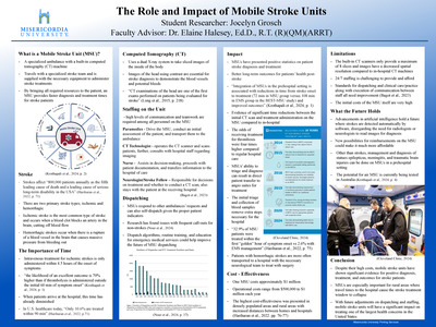
Strokes are one of the leading causes of death and disability in the United States. Optimal outcomes in stroke care depend on rapid diagnosis and immediate treatment. In hospitals, stroke protocols prioritize rapid computed tomography (CT) scans and bloodwork to diagnose and treat the patient promptly as needed. A mobile stroke unit (MSU) is a specialized ambulance equipped with a stroke team, a built-in CT scanner, and the necessary medications for treatment. This pre-hospital intervention has significantly decreased diagnosis and interventional treatment times, leading to better patient outcomes. In particular, MSUs are important for enabling the administration of treatments, such as intravenous thrombolysis, before the patient arrives at the hospital. Despite high costs and issues with staffing and dispatching, MSUs demonstrate clinical efficiency and prove to be cost-effective, especially in urban and rural areas. With ongoing advancements in technology, as well as possibilities for reimbursement, MSUs hold a promising future in the evolution of stroke care.
Keywords: stroke, mobile stroke unit, computed tomography, intravenous thrombolysis
-

HDR Brachytherapy Treatment for Basal Cell Carcinoma
Makayla L. Kester
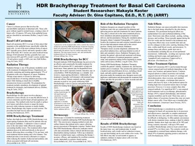
This research explains basal cell carcinoma (BCC), the most common form of skin cancer, and the role radiation therapy can play in treating patients with the disease. General information about BCC, common locations of occurrence, diagnosis methods, treatment options, side effects, and expected outcomes are discussed. BCC most often appears on sun-exposed areas such as the face, ears, and neck. For patients who are not good surgical candidates or when surgery could result in disfigurement, radiation therapy offers an effective, non-invasive alternative. One advanced technique is high-dose-rate (HDR) surface brachytherapy, which uses detailed imaging and customized applicators to deliver targeted radiation directly to the tumor. This method allows for high precision while sparing surrounding healthy tissue, making it especially useful for elderly patients or those with multiple lesions. Treatments are typically given on an outpatient basis over the course of several days. HDR brachytherapy has shown promising results in terms of local control and cosmetic outcomes. As this treatment method continues to advance, future research should focus on improving post-treatment monitoring and long-term care.
-

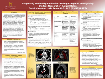
Diagnosing Pulmonary Embolism Utilizing Computed Tomography
Abigail Nolan
The purpose of this project is to explore the evolution of computed tomography (CT) technology in diagnosing a pulmonary embolism (PE) and highlight its superiority over other imaging modalities in modern medical practice. A pulmonary embolism (PE) is a life-threatening condition caused by a blood clot blocking blood flow in the lungs. It is one of the leading causes of sudden death and has severe complications. Rapidly and accurately diagnosing PE is critical for effective treatment and improved patient outcomes. Computed tomography pulmonary angiography (CTPA) has become the gold standard for diagnosing PE due to its high sensitivity, specificity, and ability to provide detailed images. The evolution of CT equipment and advancements in imaging technology has significantly improved diagnostic accuracy, scan speed, and accessibility. Compared to ultrasound (US), which is limited in its ability to directly visualize pulmonary arteries, CTPA offers a more accurate and reliable assessment.
-

Radiation Dose in Computed Tomography
Jonathan Paniagua
This poster provides detail of the radiation doses patients are exposed to in computed tomography (CT). CT helps give radiologists a more detailed image of patients. With CT first coming in the United States in the 1970s the usage has increased every decade. Presently over 100 million CT examinations are done annually. With increased usage some patients are being overexposed by scanners. A study done in the late 2000s found patients received up to eight times the radiation needed. Incidents such as this caused the need for imaging campaigns to be launched by the American Society of Radiologic Technologists to safely radiate patients. Different imaging procedures of the same area of interest can be done that cause different amounts of radiations. Different amounts of radiation to the patient can range depending on the exam. X-ray has much lower radiation doses then CT. Scanners now can still give too much radiation. Research found this can be two to three times more radiation then needed. CT is responsible for 2% of new cancer cases each year. This equals about 36,000 cases yearly.
-

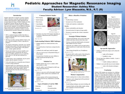
Pediatric Approaches for Magnetic Resonance Imaging
Ashley Riha
Abstract
Pediatric magnetic resonance imaging is often challenging due to the lack of cooperation and the inability to fully understand the expectations required for the examination. Children are often restless and may have difficulty remaining still for a long period of time. This can lead to motion artifacts and compensate diagnostic value. Due to these reoccurring challenges, there are several strategies to help relax the nerves of pediatric patients. Sedatives can be administered to relax the patients and assist in cooperation during the long scanning times. However, sedation safety, dosage, and patient monitoring is essential when administering medication. Alternative approaches without the use of sedation should be favored if possible. Animal-assisted therapy, musical therapy, and audio/visual recordings can act as positive distractions to help pediatric patients cope during their scan. The specific approach will be dependent on the patient’s age. Motion can also be compensated by the use of saturation bands, breathing techniques and shortened scanning sequences. With the use of these methods, MRI scans can be more tolerable for pediatric patients.
Keywords: Magnetic resonance imaging, MRI, pediatric medical imaging, pediatric sedation, Child-friendly medical environments, pediatric MRI, techniques without sedation
-

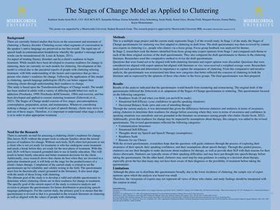
The Stages of Change Model as Applied to Cluttering: Next Steps
Kathleen Scaler Scott, Samantha Delmar, Sarah Hardy, Emma Schaedler, Erica Scheinberg, Kateri Grice, Brenna Field, Margaret Koester, Emma Maley, Kayla Montemarano, and Brooke Price
This study is based upon the Transtheoretical/Stages of Change model. The model has been studied in adults with a variety of health behaviors such as addiction (Prochaska, 1999; Prochaska &DiClemente, 1984) and has been applied to current stuttering treatment (Zebrowski et al., 2021) to assess readiness for change. The Stages of Change model consists of five stages: precontemplation, contemplation, preparation, action, and maintenance. Whenever considering making a change, as one would do as part of speech therapy, clients may move in and out of these stages. Therefore, it is important to understand what stage a client is in in order to plan appropriate treatment.
-

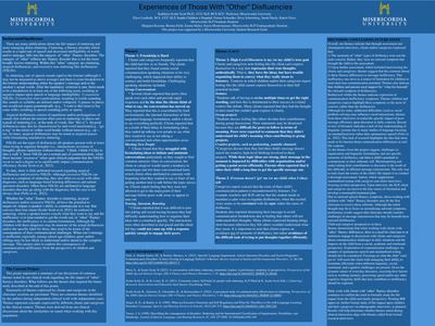
Experiences of Those With "Other" Disfluencies
Kathleen Scaler Scott, Emma Schaedler, Erica Scheinberg, Sarah Hardy, Margaret Koester, Brenna Field, Emma Maley, Kayla Montemarano, and Kateri Grice
This ethnographic study collected and analyzed interviews of 8 children with “other” fluency disorders: cluttering, atypical disfluency, excessive non-stuttering like disfluency. Each school-age child participated in a semi-structured interview focusing on grand and mini tour questions. The participants were asked to speak about their perspective and experience with communication in general. Preliminary results of this study revealed the following themes: friendships are hard; high-level communication is a goal; everyone doesn’t ‘get’ me when I talk. Clinical and research implications of findings will be discussed.
-

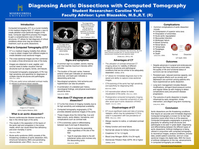
Diagnosing Aortic Dissections with Computed Tomography
Caroline York
Aortic dissections are life-threatening conditions that require an immediate and accurate diagnosis to prevent severe complications, and in some instances death. An aortic dissection is a tear that occurs within the inner layers of the aorta. Symptoms include sudden intense chest pain, shortness of breath, and stroke-like symptoms. This research discusses the utilization of computed tomography (CT) in diagnosing aortic dissections, highlighting its advantages in speed and precision through advancing technology. Computed tomography angiography (CTA) has become a vital imaging modality in the medical imaging field. CTA uses an iodinated contrast media to enhance blood vessels by making them a bright white color on the cross-sectional images. Contrast allows for clearer detection of abnormalities such as blockages, aneurysms, dissections, or other vascular pathologies. CT enables detailed visualization of the aorta and its branches through the use of 3-D reconstructions. Recent advancements in CT technology, including multi-detector and artificial intelligence (AI), further enhance diagnostic efficacy by improving image resolution and analysis. The ability to quickly and accurately diagnose aortic dissections through CT, plays a vital role in guiding timely surgical interventions and optimizing patient management, ultimately improving clinical outcomes.