

Faculty Advisor(s)
Loraine Zelna
Files
Abstract
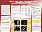
The purpose of this project is to explore the evolution of computed tomography (CT) technology in diagnosing a pulmonary embolism (PE) and highlight its superiority over other imaging modalities in modern medical practice. A pulmonary embolism (PE) is a life-threatening condition caused by a blood clot blocking blood flow in the lungs. It is one of the leading causes of sudden death and has severe complications. Rapidly and accurately diagnosing PE is critical for effective treatment and improved patient outcomes. Computed tomography pulmonary angiography (CTPA) has become the gold standard for diagnosing PE due to its high sensitivity, specificity, and ability to provide detailed images. The evolution of CT equipment and advancements in imaging technology has significantly improved diagnostic accuracy, scan speed, and accessibility. Compared to ultrasound (US), which is limited in its ability to directly visualize pulmonary arteries, CTPA offers a more accurate and reliable assessment.
Publication Date
2025
Document Type
Poster
Department
Medical Imaging
Keywords
pulmonary embolism, PE, computed tomography, CT, pulmonary angiography, CTPA, ultrasound, US
Disciplines
Medical Sciences | Medicine and Health Sciences
Recommended Citation
Nolan, Abigail, "Diagnosing Pulmonary Embolism Utilizing Computed Tomography" (2025). Student Research Poster Presentations 2025. 5.
https://digitalcommons.misericordia.edu/research_posters2025/5