

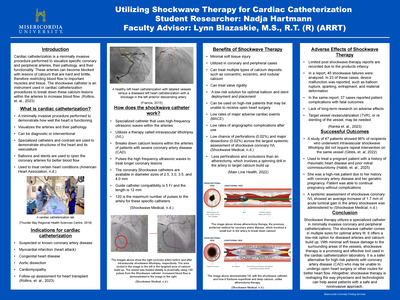
Medical Imaging Senior Posters showcase innovative research by Misericordia University Medical Imaging winter graduates.
-

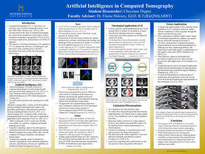
An Examination of Arterial Closure Devices
Colin Hart
The intention of this poster is to provide information on the various devices and methods for achieving hemostasis after arterial access has been used for cardiac catheterization procedures. There are a multitude of closure devices being utilized to stop patient bleeding following a cardiac catheterization procedure, and keeping up with the information and data related to these devices can be complex. Suture based, collagen based, patch based, membrane based, pressure bands, as well as application of manual pressure are some of the devices and/or methods that will be examined within this project. A thorough overview of these closure devices currently being used within cath labs across the globe will be compared and contrasted. Some cardiac procedures that would require femoral and radial artery access will be listed, as well as a brief explanation as to why these access sites are preferred. Information and statistics on the usage, success rates, as well as likelihood of complications for each device/method will be included in the slides. Details regarding the specifics of complications that may occur will also be explained. Ultimately, this poster seeks to inform viewers about the plethora of options available to avoid post procedure bleeding should they ever find themselves in need of cardiac intervention, and to be able to discuss with their doctors what the safest of these options may be.
Keywords: Cardiac Catheterization, Arterial Closure, Complications Associated With Arterial Closure, Hemostasis, Sutures for Arterial Closure, Collagen-based Arterial Closure, Patch-based Arterial Closure, Manual Arterial Pressure, Membrane-based Closure, Arterial Pressure Bands
-

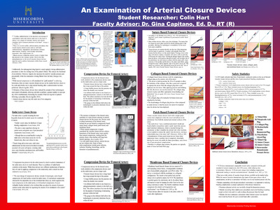
SGRT Eliminates Need for Patient Tattoos in Radiation Therapy
Gina Hemsley
Surface Guided Radiation Therapy, or SGRT, is a rapidly growing technology designed to improve radiation treatment accuracy of nearly every cancer type without the need for permanent skin tattoos. Patients were typically marked with 3 tattoos, indicating patient setup marks and isocenter marks. SGRT is an external beam radiation therapy technique which uses three-dimensional camera technology to accurately target and kill cancer cells. During patient setup and treatment, the technology uses the patient’s external surface to guarantee that the radiation dose is being consistently applied in accordance with the treatment plan, as well as tracking any patient motion throughout treatment. The real-time positioning tracking acts as a safeguard during radiation treatment, immediately stopping if the patient moves out of exact position. Unlike other forms of radiation therapy, SGRT ensures the patient is receiving radiation therapy with submillimeter accuracy without the need for permanent skin tattoos, as well as shorter treatment times, greater protection of healthy tissues, and higher patient comfort.
-

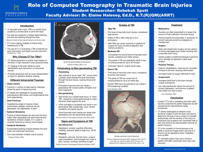
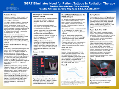
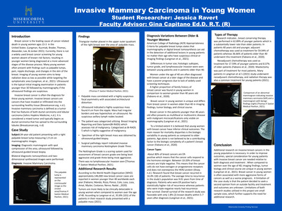
Subdural Hematomas and the Help of Computed Tomography
Kelsey Jackson
Subdural hematomas can occur at any age but are most commonly seen in the elderly population. Subdural hematomas affects 21 in every 100,000 individuals, and are becoming more common. They can occur for a variety of reasons such as trauma or underlying health conditions. Subdural hematomas occur due to a collection of blood in the subdural space between the dura and arachnoid mater. Computed tomography is the best imaging modality in the aiding of diagnosing subdural hematomas due to the easy accessibility, short scan times, and the ability to create cross sectional images. Subdural hematomas can be classified into categories based on the location and appearance of the bleed. These classifications can include acute, subacute, and chronic. Dependent on the location and diagnosis of the subdural hematoma non-surgical intervention such as monitoring, or medication may be needed. Surgical intervention may be needed based on the severity of the bleed and its classification. If a subdural hematoma is caught in a timely manner with successful intervention there is a lower chance of reoccurrence, there is a 3% to 20% post-operative reoccurrence rate. The overall prognosis is patient dependent and considers the size and classification of the bleed.
-

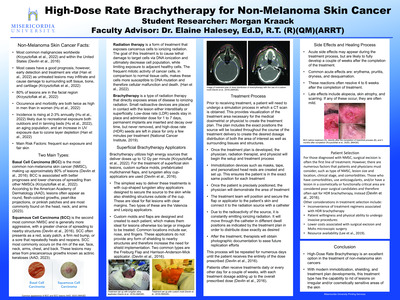
Detecting a Pulmonary Embolism Using Computed Tomography
Marissa O'Brien
Abstract
A pulmonary embolism is a potentially fatal disease in which a blood clot obstructs the pulmonary artery. Pulmonary emboli affect thousands of people every year in the United States. A timely diagnosis is vital to ensure quick and proper treatment and resolution. Signs and symptoms can vary among different patients, but some include shortness of breath, chest pain, syncope, and leg pain. Computed tomography (CT) is the golden standard for diagnosis, specifically a CT pulmonary angiogram, which quickly and accurately visualizes clots in the pulmonary artery using iodinated contrast in the form of a bolus better than any other modality. Other CT studies may be performed such as a non-contrast chest scan or performance of the study in the venous phase. Treatments vary depending on the severity of the emboli and range from medications to surgery. With advancements in medicine and diagnostic tests, mortality rates have significantly decreased. The prognosis in patients with pulmonary emboli have improved with timely intervention.
-

Surface Guided Radiation Therapy versus Traditional Triangular Positioning
Katelyn Sausser
Surface-guided Radiation Therapy (SGRT) is a positioning monitoring system utilizing 3D nonionizing optical surface imaging. This system assists in patient set-up and allows for real-time monitoring of the patient’s skin surface during radiation therapy treatment. Surface-guided imaging can be used for many treatment sites such as breast, abdomen, head and neck, and extremities. SGRT is believed to have more advantages than the traditional triangular positioning including shorter set-up times, improved accuracy and reproducibility of treatment area, decreased dose, and improved patient comfort. Studies have also shown the planned dose delivery can also be monitored with SGRT and decrease dose to surrounding tissues.
-

MRI Guided NeuroBlate Laser Ablations
Sarah Bielfeld
This project explains how magnetic resonance imaging (MRI) is used for neuroblate laser ablations, or laser interstitial thermal therapy (LITT). LITT therapy involves the usage of heat to treat intracranial pathologies such as glioblastomas. Laser therapy is an non-invasive alternative to open resection and craniotomy procedures in which a surgical opening of the skull and brain tissue are necessary. Ablations are considered a safer alternative because patients showed no signs of declining cognitive functions post surgery. Magnetic resonance scanners are used in conjunction with laser systems. The two types of systems are Visualase (Visualase, INC.) and NeuroBlate (Monteris Medical, Inc.) that demonstrates the temperature of surrounding tissue in both the central and peripheral zones of the brain. Patient is under general anesthesia while a “burr” hole is drilled into their head for entry of the laser probes. MRI uses T1 sequences to demonstrate the full length of the probe and FLAIR sequences are utilized for anatomical reference. The ablated lesion can be seen in the axial view of a MR image; then diminishes in size up to three months.
Keywords: Magnetic Resonance Imaging (MRI), Neuroblate Laser Ablation, Laser Interstitial Therapy (LITT), Visualase, NeuroBlate, Glioblastoma
-
 by Ashlyn Case")
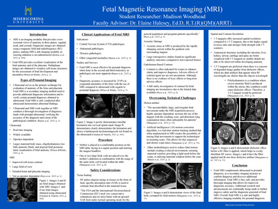
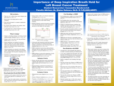
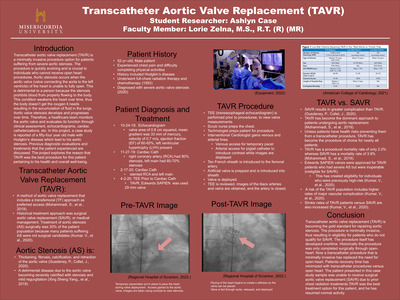
Transcatheter Aortic Valve Replacement (TAVR)
Ashlyn Case
Title of Poster: Transcatheter aortic valve replacement (TAVR) Student Researcher: Ashlyn Case Faculty Advisor: Loraine D. Zelna, M.S., R.T. (R)(MR) Internship Mentor: Stephanie Jugus, B.S., R.T., RCIS Internship Site: Cardiac Cath, Regional Hospital of Scranton, Scranton, PA Abstract Transcatheter aortic valve replacement
(TAVR) is a minimally invasive procedure option for patients suffering from severe aortic stenosis. The procedure is quickly evolving and is crucial to individuals who cannot receive open heart procedures. Aortic stenosis occurs when the aortic valve (valve connecting the aorta to the left ventricle) of the heart is unable to fully open. This is detrimental to a person because the stenosis prohibits blood from properly flowing to the body. This condition weakens the heart over time, thus the body doesn’t get the oxygen it needs resulting in the accumulation of fluid in the lungs. Aortic valve stenosis develops and progresses over time. Therefore, a healthcare team monitors the aortic valve and evaluates its function through clinical assessment, echocardiograms, cardiac catheterizations, etc. In this project, a case study is reported of a fifty-four-year-old male with Hodgkin’s disease which lead to his aortic stenosis. Previous diagnostic evaluations and treatments that the patient experienced are discussed. The project explores the reason that TAVR was the best procedure for this patient pertaining to his health and overall well-being.
Keywords:Transcatheter, aortic stenosis, evolving, minimally invasive
-
 by Hailey Clark")
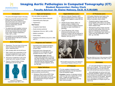
Imaging Aortic Pathologies in Computed Tomography (CT)
Hailey Clark
Computed tomography (CT) is an increasingly important imaging modality to visualization of aortic pathologies. The aorta is the largest vessel in the body and gives rise to several important arteries that supply surrounding organs, so pathologies of this vessel can prove dangerous. The two main pathologies that affect the aorta are aneurysms and dissections, each of which can rupture and quickly become fatal. Symptoms for these pathologies can vary widely but primarily include pain localized to the area of the aorta that is affected, with risk factors encompassing several heart conditions as well as genetic factors. The prognosis for aortic conditions has improved as a result of CT technology but is still relatively grim, with treatment options including invasive and minimally invasive surgical interventions along with medicinal management and screening for the rest of the patient’s life. CT provides several options for improving visualization of the pathology including valuable post-processing and reconstruction images that can maximize detail and allow for quicker and easier diagnoses, improving patient prognosis, and the use of ECG gating to virtually eliminate motion artifact from the heart that can obscure or mimic pathology. The continuous advancements in CT technology are only becoming more readily available, creating an expectation that the prognosis for these serious conditions will continue to become more favorable.
Keywords: Computed Tomography, Aorta, Dissection, Aneurysm, Post-Processing, Image Reconstruction, ECG Gating
-
 for Diagnosis of Parkinson’s Disease by Samantha Clark")
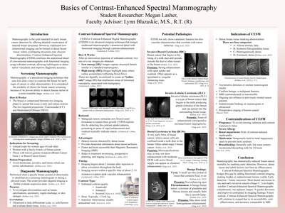
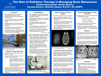
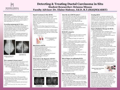
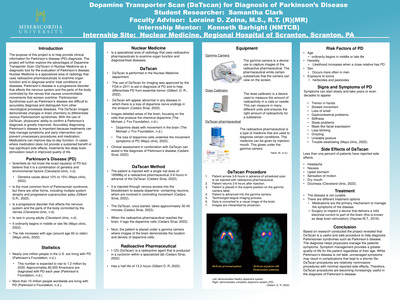
Dopamine Transporter Scan (DaTscan) for Diagnosis of Parkinson’s Disease
Samantha Clark
The purpose of this project is to help provide clinical information for Parkinson’s disease (PD) diagnosis. The project will further explore the advantages of Dopamine Transporter Scan (DaTscan) in Nuclear Medicine as a diagnostic tool for the evaluation of Parkinson’s disease. Nuclear Medicine is a specialized area of radiology that uses radioactive pharmaceuticals to examine organ function and to diagnose and/or treat conditions or diseases. Parkinson’s disease is a progressive disorder that affects the nervous system and the parts of the body controlled by the nerves that causes uncontrollable movements that worsen overtime. Parkinsonian Syndromes such as Parkinson’s disease are difficult to accurately diagnose and distinguish from other neurological processes diseases. The DaTscan images demonstrate changes in brain chemistry to differentiate various Parkinsonism syndromes. With the use of DaTscan, physicians' ability to confirm a Parkinson’s diagnosis is greatly improved. Accurately diagnosing Parkinson’s disease is important because treatments can help manage symptoms and early intervention can prevent unnecessary procedures and medication. Medications can improve day-to-day function. In cases where medication does not provide a sustained benefit or has significant side effects, treatments like deep brain stimulation result in improved quality of life.
-

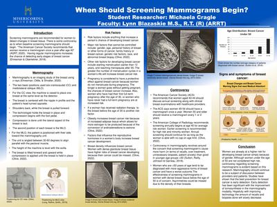
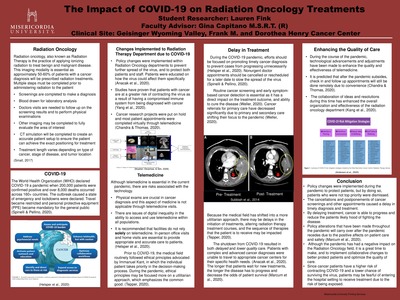
When Should Screening Mammograms Begin?
Michaela Cragle and Lynn Blazaskie
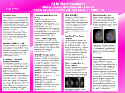
When Should Screening Mammograms Begin?
Michaela Cragle
Lynn Blazaskie M.S., R.T. (R) (ARRT)
Abstract
Screening mammograms are imaging exams performed yearly to evaluate breast tissue. As women age, changes in breast tissue can be normal or can be an early sign of cancer. The age women should begin screening mammograms is not clear. According to literature it has created much controversy over the years. Most women begin the screening process at the age of 40, and even earlier depending on family history. Research groups from multiple institutions followed and evaluated women who received yearly mammograms. Most women were divided into groups based on age to see if recalls, breast cancer, or biopsies were more common in specific age groups. Some would say that the amount of recalls and biopsies that happen are excessive, and are being too cautious. Results showed that women of younger ages tend to get called back more for benign findings rather than women who are older. This research demonstrates that if cancer can be detected at an earlier age, the outcome for the patient is better. Being a little more cautious is better than overlooking something that could potentially become worse. The age at which screening mammograms should begin may change in the future as technology advances and research is ongoing.
Keywords: screening mammogram, recall, benign, cancer, biopsy, age, controversy, family history, early
-

The Visualization of Abdominal Aortic Aneurysms with the Aid of Computed Tomography
Graham Dileo
The Visualization of Abdominal Aortic Aneurysms with the Aid of Computed Tomography
Graham J. Dileo
Dr. Gina Capitano Ed.D., R.T. (R)
Geisinger Community Medical Center, Scranton, PA
Abstract
This research discusses Abdominal Aortic Aneurysms (AAA) and the role of Computed Tomography (CT) in diagnosing and establishing a treatment plan. An AAA is a bulge or swelling in the descending aorta, the primary blood vessel in the human body, that can be life-threatening. This type of aortic aneurysm is one of the most common causes of death for patients with a history of high blood pressure or heavy smoking. AAAs typically occur in white men, ages 65 to 75. However, are not limited to this population as the research represents a case study of a 74-year-old female diagnosed with an AAA. Since AAAs can be deadly due to possible rupture, obtaining quick yet qualitative diagnostic imaging is possible with CT. This type of imaging provides cross-sectional imaging of the human body, which can be vital in assessing the size and shape of an AAA. Early detection of an AAA will determine the proper route of treatment. Smaller-sized AAAs of 1-5 cm typically require medication and close monitoring, while larger-sized AAAs greater than 6 cm require emergency surgery. These surgeries are typically endovascular. Although the mortality rate is high, CT can help determine the correct actions needed to prevent possible rupture and increase chances of survival.
Keywords: rupture, computed tomography, abdominal aortic aneurysm, risks, surgery
-
 for Treatment of Lung Cancer by Camryn Frazier")
Stereotactic Body Radiation Therapy (SBRT) for Treatment of Lung Cancer
Camryn Frazier
This project explains stereotactic body radiation therapy (SBRT) and its role in treating lung cancers. Different types of SBRT treatment, statistics, toxicity, success rates, and data from studies are discussed. More than half of all cancer patients experience lung metastasis, many of these cases being inoperable. SBRT provides a noninvasive alternative to surgery as well as high rates of local tumor control and minimal toxicity. SBRT is a form of external beam radiation therapy that delivers a hypofractionated dose directly to the cancerous target volume. Two common types of SBRT utilized to treat lung cancer are intensity modulated radiation therapy (IMRT) and volumetric modulated arc therapy (VMAT). Patients with lung metastasis have demonstrated very low toxicity from SBRT, with most patients reporting below stage 3. 10-15% of early stage non-small cell lung cancer cases treated with SBRT result in local recurrence. Salvage SBRT treatment of the lungs has shown to be mostly successful with an overall survival rate of 68%. Due to the amount of SBRT treatment cases resulting in local recurrence resulting in the need for salvage SBRT treatment, there is a need for continued research to reduce the statistic.
-
 by Emily Paciga")
Use of CT to Diagnose Subcutaneous Emphysema (SE)
Emily Paciga
Subcutaneous emphysema is a follow up symptom to a traumatic event, surgery, or nose-blowing. In addition subcutaneous emphysema is considered benign and self limiting. Computed tomography (CT) is the imaging modality of choice for diagnosing subcutaneous emphysema. CT is preferred because of its sensitivity to tissue densities. A CT will demonstrate dark air pockets within the subcutaneous layer of the skin. Furthermore, upon palpation of the the skin a crepitus crackling sound is demonstrated. Moreover, listening to the skin with a stethoscope will emit a high-frequency acoustic sound. Nevertheless, extensive subcutaneous emphysema may require medical assistance. Typically, orbital subcutaneous emphysema is non-life threatening and will resolve within a few days as the body absorbs the air. If the air is extensive and compresses on the optic nerve medical attention is required to prevent vision loss. In addition, traumatic subcutaneous emphysema may be caused from pneumothorax or other blunt trauma. The air rises and spreads within the body and can travel though different anatomical planes. Lastly, subcutaneous emphysema may be a complication of surgical procedures. Individuals are susceptible to suffering from subcutaneous emphysema after procedures like colonoscopies. To conclude, subcutaneous emphysema is a symptom that can be diagnosed by CT and is relatively harmless.
Keywords: Subcutaneous emphysema, CT, benign, crepitus
-

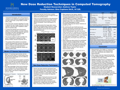
Stereotactic Body Radiation Therapy for Spinal Metastases
Kendall Pearage
SBRT for Spinal Metastases
Kendall Pearage
Dr. Gina Capitano Ed. D, R.T. (R)
Cathy Moody R.T. (T)
Northeast Radiation Oncology Center
Abstract
Spinal metastases are the most common spinal tumors accounting for approximately 90% of cases and are a result of cancer arising from another part of the body. This research explains how Stereotactic Body Radiation Therapy (SBRT) can treat spinal metastases. SBRT is a type of treatment that uses high fractionated doses of radiation to treat patients with different cancers. SBRT is commonly used to treat spinal metastases because it allows radiation therapists to treat a very precise location on the spine, causing little damage to nearby structures and organs. Patients typically receive one to five fractions of treatment, each with varying doses. The radiation targets and eventually shrinks the tumor bed while administering little dose to the spinal cord. SBRT for spinal metastases has shown longer survival rates and increased pain relief in patients. Continuing research is needed to determine any negative effects Stereotactic Body Radiation Therapy can cause to cancers that spread to bone.
Keywords: SBRT, Spinal Metastases, Cancer, Radiation, Treatment, Spinal Cord
-

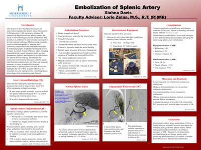
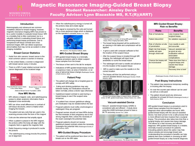
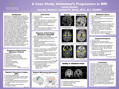
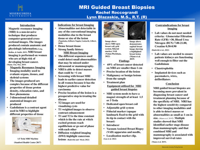
MRI Guided Breast Biopsy
Rachel Roccograndi
Magnetic resonance imaging (MRI) is a non-invasive technique that produces computer-generated cross-sectional images. The images produced contain anatomic and physiologic information. Using non-ionizing radiation, MRI creates images through the interaction of magnetic fields and radio frequency energy on biologic tissue. The specificity present in the images produced is the main reason MRI is a popular choice of diagnostic testing. MRI is used to evaluate organs, tissues, and skeletal systems and different sequences are used depending on what the main focus of the test is. MRI images are more complex because they contain information about different properties of tissue-proton density, relaxation rates, and flow phenomena. MRI breast imaging is performed on women who are at high risk of developing breast cancer, or dense breasts because it is a highly detailed test. The different sequences used could detect small abnormalities that may be missed under ultrasound or mammography. MRI guided breast biopsies are done when an abnormality is detected and a preoperative step in treating the disease. The specialized equipment is helpful in determining the exact size, location, and extent of the cancer before choosing a path of treatment. MRI guided breast biopsies are becoming more prevalent in diagnosing and planning treatment for women who have been diagnosed with breast cancer because of the specificity.
Keywords: Magnetic resonance imaging, non-ionizing radiation, MRI breast imaging, MRI guided breast biopsy
-

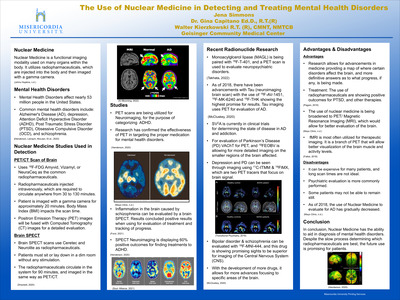
The Use of Nuclear Medicine in Detecting and Treating Mental Health Diseases
Jena Simmons, Gina Capitano, and Walter Kierzkowski
Mental health disorders affect over 792 million people around the world, and a leading cause for disability. Mental health disorders include a plethora of conditions that affect mood, thinking and behavior, including but not limited to, depression, dementia, anxiety, autism, and schizophrenia. The most common method of diagnosis is a psychological evaluation, which can lack accuracy if performed as the only method of diagnosis. Nuclear Medicine helps aid in diagnosis of these disorders in conjunction with psychological testing. Brain scans can help determine if there is a pathogenic cause for the symptoms. Brain scans are done utilizing Positron Emission Tomography (PET) scans and Single Photon Emission Computerized Tomography (SPECT) scans. Advancements in these studies allow Nuclear Medicine to better aid in diagnosis and treatments of this wide range of disorders.
Keywords: Nuclear Medicine, PET, SPECT, mental illness, diagnose, treatment, mental health.
-

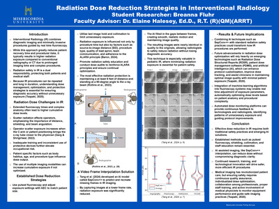
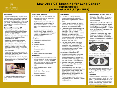
Low Dose CT Scanning for Lung Cancer
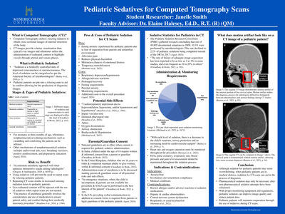
Patrick Strouse
Low Dose CT scanning for Lung Cancer
Patrick Strouse
Lynn Blazaskie M.S.,R.T.(R)(ARRT)
Abstract
Computed Tomography (CT) is the imaging of patients in a cross-sectional plane using x-ray. CT is used to diagnose diseases and is producing more efficient ways of this by making radiation dose lower, this leads to Low Dose CT and its abilities in diagnosing lung cancer. The research portrayed in this project provides points across the different aspects of Low Dose Computed Tomography (CT) and its relation to lung cancer to explain the growing new scan of Low Dose CT. Lung cancer is described as a cancer originating in the lungs which the cells grow exponentially causing many mortalities in the world. Lung cancer can be determined by many risk factors such as smoking, radon exposure, family history of lung cancer, and even diet. The symptoms for lung cancer may not be apparent early on, but many researchers find that detecting it early is the best chance for survival. Incorporating evidence shows that Low Dose CT is the best way to detect lung cancer early and to substantially raise the survival rate of this deadly disease, in evidence it shows the statistics of early findings on these scans as compared to others. This research also brings into consideration some of the reasons people may not have access to these scans, this is because of the newer technology and many do not know of it or its effectiveness to save lives. Some future work with these findings should raise the awareness of this scan and educate more individuals.
Keywords: Lung Cancer, Low Dose CT, Computed Tomography, Health
-
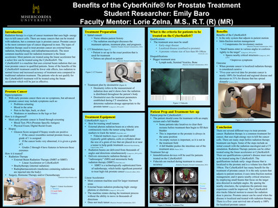
 for Gastric Varices (GV) in Interventional Radiology (IR) by Lauren Wiest")
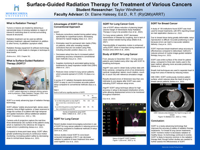
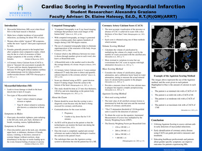
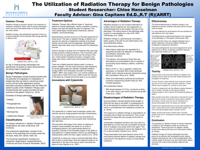
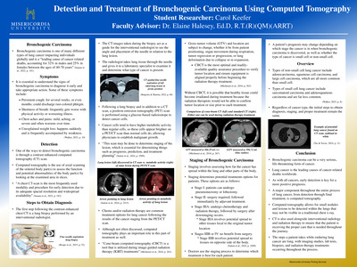
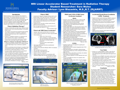
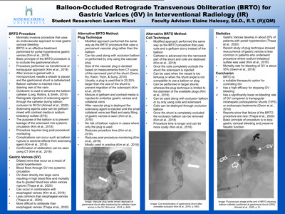
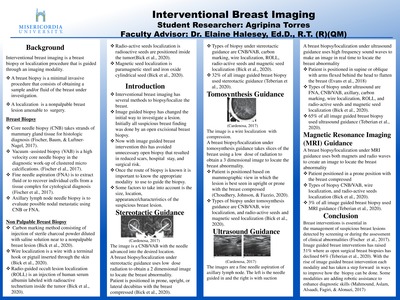
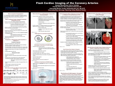
Balloon-Occluded Retrograde Transvenous Obliteration (BRTO) for Gastric Varices (GV) in Interventional Radiology (IR)
Lauren Wiest
Balloon-Occluded Retrograde Transvenous Obliteration (BRTO) for Gastric Varices in Interventional Radiology (IR)
Lauren Wiest
Dr. Elaine Halesey, Ed.D., R. T. (R) (QM)
Abstract
This project explains gastric varices and the role the BRTO procedure has in treating gastric variceal bleeding. Statistics regarding gastric varices and the modified techniques of the procedure are discussed. Gastric varices are dilated veins that can rupture at any time and the main cause of gastric varices is portal hypertension. Gastric varices develop in approximately 20% of patients with portal hypertension. Portal hypertension is the result of liver disease or cirrhosis. The BRTO procedure has the potential to help patients suffering from gastric varices by obliterating these varices before they rupture or while their bleeding to save patients’ lives. This procedure can be performed using just an occlusion balloon, a plug, or coils to occlude the shunt. In some cases, doctors might use more than one technique. BRTO is an effective procedure used to treat gastric varices. Reports show that failure of the BRTO are rare. This minimally invasive procedure has high success rates and minimal complications. The high success rate of this procedure demonstrates how valuable this procedure really is.
Keywords: Balloon-Occluded Retrograde Transvenous Obliteration (BRTO), Gastric Varices, Interventional Radiology (IR), Portal Hypertension
-

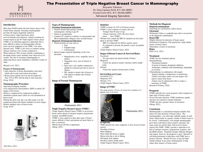
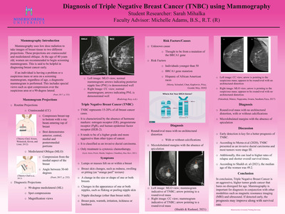
The Presentation of Triple Negative Breast Cancer in Mammography
Reayanne C. Yokimcus, Gina Capitano, and Crystal Crawn
Triple Negative Breast Cancer (TNBC) is a type of breast cancer absent of estrogen and progesterone within the cancer cells. Additionally, TNBC is absent of a large amount of the protein HER2. Negative testing for all three elements is indicative of TNBC. Approximately 10-15% of women with breast cancer are diagnosed with TNBC. African American women under 40 years of age and women with the BRCA1 mutation are most commonly diagnosed with TNP. The BRCA1 gene is important when fighting off cancer. If there is a mutation within the BRCA1 gene, then that makes men and women more susceptible to cancer. Although rare, TNBC is extremely invasive and typically has a poor prognosis. Symptoms of TNBC include swelling, pain, nipple discharge, swollen lymph nodes, and a hard lump within the breast. It can be diagnosed through different imaging modalities and biopsies. Treatment includes surgery, chemotherapy, and radiation therapy. There are different stages of TNBC that require different treatments.
Keywords: Triple Negative Breast Cancer, TNBC, Breast Cancer Treatment
-

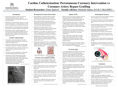
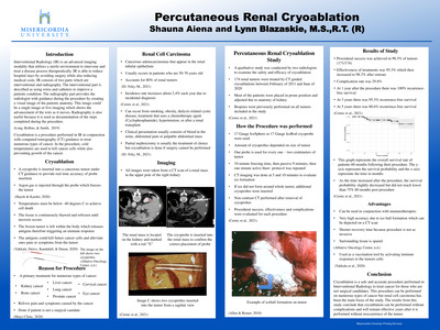
Percutaneous Renal Cryoablation
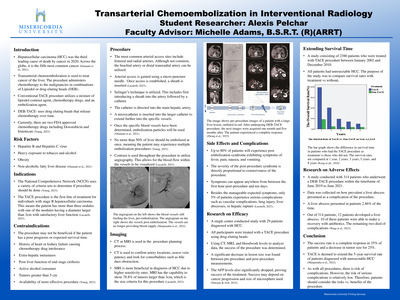
Shauna M. Aiena
This research topic discusses how safe and effective the use of cryoablation is to treat a renal tumor. Cryoablation is a procedure performed in the Interventional Radiology department to kill cancer cells using very cold temperatures. This procedure is done when the patient cannot have surgery to remove the tumor itself. Although the tumor is not removed during cryoablation, the treatment is performed to freeze the tumor to prohibit growth while releasing antigens from the tumor which triggers an immune response. To see if cryoablation is as effective as some say, a qualitative study was conducted which used 174 renal tumors that were treated by computed tomography (CT) guided cryoablations done between February of 2011 and June of 2020. Procedural success, effectiveness and complications were evaluated for each procedure. The results of this study were that in 98.3% of tumors (171/174), procedural success was achieved. The effectiveness of treatments was 95.3% which then increased to 98.2% after retreats. Overall, there was a complication rate of 29.8%. At 1 year after the procedure there was 100% recurrence-free survival, at 3 years there was 95.3% recurrence-free survival and at 5 years there was 88.6% recurrence-free survival. From this study one can conclude that cryoablation is a safe and effective procedure even years after it is performed. This technique is able to treat small renal tumors without any major complications. Possible implications from this research could be used while working in the interventional radiology department to ensure patients understand that this procedure is safe and effective.
Keywords: cryoablation; renal tumor treatment; renal cryoablation; interventional radiology
-
 by Ashlee Blannett")
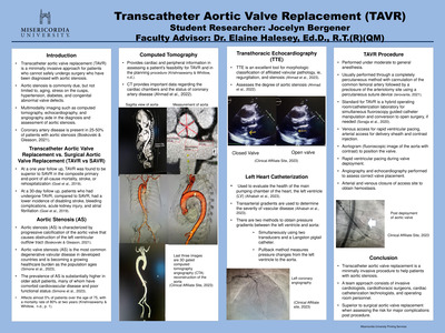
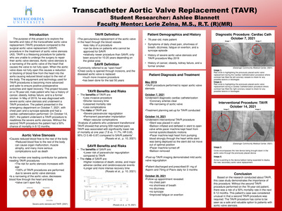
Transcatheter Aortic Valve Replacement (TAVR)
Ashlee Blannett
Transcatheter Aortic Valve Replacement Versus Surgical Aortic Valve Replacement in Treatment of Aortic Valve Stenosis
Ashlee Blannett
Faculty Mentor: Lorie Zelna, M.S., R.T. (R)(MR)
Abstract
The purpose of this project is to explore the benefits and risks of the transcatheter aortic valve replacement (TAVR) procedure compared to the surgical aortic valve replacement (SAVR) procedure in the treatment of aortic valve stenosis (AS). TAVR is a lifesaving procedure for patients who are unable to undergo the surgery to fix their aortic valve stenosis. Aortic valve stenosis is a narrowing of the aortic valve of the heart that causes the valve to not open fully. When the aortic valve does not open fully this causes a reduction or blocking of blood flow from the heart into the aorta. The equipment and technology used for TAVR procedures is becoming more advanced and safer to ensure postoperative outcomes are positive and recovery is quick. This project focuses on a male, 78-year-old, patient that underwent a TAVR procedure on October 14, 2021. The patient was diagnosed with severe aortic valve stenosis and had a TAVR procedure done previously in May of 2019. There was a history of cancer, obesity, kidney failure, and being a former smoker. Initial visit to the hospital’s emergency room was chest pain and a syncope episode. Without another TAVR procedure the patient had a 50% chance of mortality in 6-12 months. Keywords: aortic valve stenosis (AS), transcatheter aortic valve replacement (TAVR), surgical aortic valve replacement (SAVR)
-

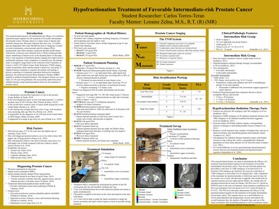
Low-Dose Lung Cancer Screening Computed Tomography
Makalie Blazick
Abstract
Low dose Computed Tomography (CT) screening is essential when finding and treating suspicious lung nodules. A CT machine uses a highly advanced detector assembly that measures the amount of radiation exiting the patient and records data. A CT machine also conducts three-dimensional reconstructions of images that is used for surgical planning, CT angiography (CTA), radiation therapy planning, and virtual reality imaging. The low dose lung cancer screening is usually reserved for older patients with the greatest risk of lung cancer, including former or current smokers. To determine if someone is eligible for low dose CT screening, the pack years are calculated by multiplying the number of packs of cigarettes smoked per day and the number of years someone has been smoking. Researchers conducted a study using 50 milliamperes-second (mAs) for a low dose CT screening instead of 150 mAs for a standard dose CT screening. The results reveal that in comparison, the low dose and standard dose offer the same diagnostic performance and characterization capabilities when searching for lung cancers. Unfortunately, low dose lung cancer screening is heavily underutilized at a rate of only 2% in the United States. Discussing the risks and benefits from a low dose CT screening is highly recommended, especially for people around the age 70.
-

MRI-Guided Breast Biopsy
Makayla Franko
This project explains the role MRI has in performing breast biopsies. MR imaging is used along with mammogram and ultrasound to detect and monitor lesions. Traditional imaging of the breast is explained to understand how images of the breast are obtained. The grade of the lesion and how the lesion is found indicates the type of biopsy performed. MRI provides superior visibility for lesions within the breast. MRI-guided breast biopsies are indicated in patients with lesions only visible on MRI. The equipment required for an MRI-guided breast biopsy includes the breast coil, grid, and vacuum-assisted breast biopsy apparatus. Experienced breast radiologists and MR technologists identify the lesion to be biopsied on axial images of the breast using special computer programming. A case study is also included within the research to provide a better understanding of a patient’s experience with MRI-guided breast biopsy. Following the biopsy, the patient receives a soft compression mammogram and has follow-up MR imaging of the breast. A summary of new technology is also included. The new technology would lead to future research that compares different MRI procedures of the breast.
Keywords: MRI, MRI-guided breast biopsy, breast lesions, case study

