

Student Research Poster Presentations showcase innovative research by Misericordia students across all disciplines.
-

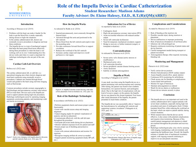
Role of the Impella Device in Cardiac Catheterization
Madison Adams
The Impella device is a form of temporary mechanical circulatory support increasingly used in the cardiac catheterization lab to assist patients undergoing high-risk procedures or experiencing severe cardiac dysfunction. This project explores how the Impella functions, when it is to be used, and the role of the radiologic technologist in supporting these procedures. The device is inserted percutaneously, most commonly through the femoral artery, and works by drawing blood from the left ventricle and expelling it into the ascending aorta. Therefore, the device is improving cardiac output and reducing the myocardial workload in total. Careful patient selection, monitoring, and management are essential due to risks such as bleeding, vascular injury, and hemolysis. Radiologic technologists play a critical role in Impella usage by ensuring proper imaging, maintaining radiation safety, assisting with device placement, and supporting overall workflow in the lab. This research emphasizes the importance of teamwork and technical skills in achieving beneficial patient outcomes. As technology continues to advance and use expands in complex cardiac cases, ongoing education and training for imaging professionals becomes increasingly important.
-

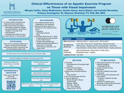
Clinical Effectiveness of an Aquatic Exercise Program on Those with Visual Impairment
Mikayla Calitis, Haley McMenimen, Isabella Bernitsky, Amelia Gansz, and Karol Bialek
-

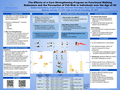
The Effects of a Core Strengthening Program on Functional Walking Endurance and the Perception of Fall Risk in Individuals over the Age of 65
Melissa Cencetti, Nicole Evanosky, Amber Hackenberg, Angela Shander, Nick Dworetsky, Ryan Zuch, and Andrew Layne
-

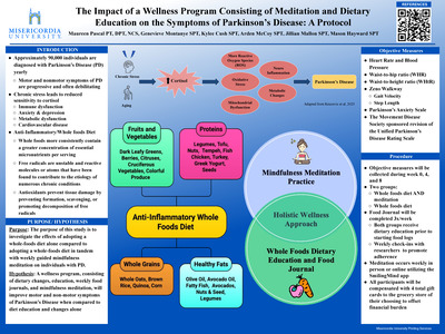
The Impact of a Wellness Program Consisting of Meditation and Dietary Education on the Symptoms of Parkinson’s Disease: A Protocol
Kylee Cush, Maureen Romanow Pascal, Genevieve Montanye, Arden McCoy, Jillian Mallon, and Mason Hayward
Individuals with Parkinson’s Disease (PD) often experience many motor and nonmotor symptoms that impact their ability to participate in their daily life. This protocol aims to address the symptoms of PD by utilizing complementary strategies to manage chronic stress and unhealthy dietary lifestyles, both of which have been identified as risk factors for PD. This eight-week mixed-design randomized controlled trial will take place at Misericordia University, Rock Steady Boxing Old Forge, and the University of Scranton, and will utilize convenience sampling through flyer outreach. Individuals will be randomized into two groups: group one will complete a whole foods diet as well as mindfulness meditation weekly, and group two will complete solely a whole foods diet. Outcome measures will be measured at week 0,4, and 8, while food journals and check-ins by researchers will occur during weeks 2, 3, 5, 6, and 7. Data collection will be processed utilizing a mixed ANOVA and the Mann-Whitney U once the study is completed. Participants will be compensated to offset the financial impact of dietary modifications.
-

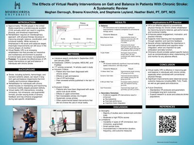
The Effects of Virtual Reality Interventions on Gait and Balance In Patients With Chronic Stroke: A Systematic Review
Meghan Darrough, Stephanie Layland, and Breena Kravchick
Background: Chronic stroke survivors often experience long-term gait and balance impairments that impact their functional abilities and activities of daily living. Emerging interventions, such as virtual reality (VR), are being evaluated on their potential to enhance rehabilitation outcomes.
Objective: This systematic review aimed to evaluate the effects of virtual reality interventions on gait and balance in individuals with chronic stroke.
Methods: Two literature searches were performed in September 2025 and January 2026 using the CINAHL Complete, MEDLINE, and PubMed databases.
Results: Fourteen studies met the inclusion criteria and were categorized based on balance outcome measures (Berg Balance Scale, Tinetti Performance-Oriented Mobility Assessment, and Timed Up and Go) and gait outcomes (10-Meter Walk Test, 6-Minute Walk Test, Dynamic Gait Index, and gait parameters). Several studies demonstrated statistically significant improvements in both balance and gait compared to conventional rehabilitation. However, variability in the type of VR intervention used was observed, and some studies reported no significant differences in outcomes.
Conclusion: Virtual reality interventions appear to be effective when used in addition to conventional physical therapy for improving balance and gait in individuals with chronic stroke. However, further high-quality research is needed to standardize protocols for virtual reality interventions and determine the effectiveness of different types of VR interventions.
-

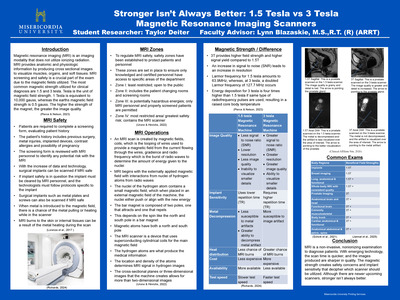
Stronger Isn't Always Better: 1.5 Tesla vs 3 Tesla Magnetic Resonance Imaging Scanners
Taylor Deiter
Abstract
Magnetic resonance imaging (MRI) is an imaging modality that does not use ionizing radiation. Instead, MRI uses magnetic fields, gradience, coils, and radio frequency to provide anatomic and physiologic information. The purpose of the research study is to examine the strengths and weaknesses of a 1.5 tesla to a 3-tesla magnetic resonance imaging scanner. Cross sectional images are produced to better visualize muscles, organs, and soft tissues for diagnostic purposes. MRI screening and safety is a crucial part of the modality. The most common magnetic strength used for clinical diagnosis are 1.5 and 3 teslas. The higher the strength of the magnet, the greater the image quality and with emerging technology, the scan time is quicker, and the images produced are sharper in quality. However, higher magnetic strengths create safety concerns and implant sensitivity that require a lower field strength such as the 1.5 tesla scanner; therefore, screening and patients’ safety is crucial. An increase in magnetic strength causes an increase in energy deposition, raising the bodies core temperature. The increase in energy deposition is what causes implant sensitivity, meaning not every patient is suitable on the higher tesla scanner. Therefore, there is still a need for the 1.5 tesla scanners over the higher strength magnets. This proves that stronger isn’t always better.
Keywords: 1.5 tesla, 3 tesla, MRI scanners, safety, magnetic strength
-

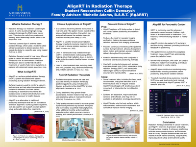
AlignRT in Radiation Therapy
Collin N. Domozych
This project focuses on the implementation of AlignRT, a surface-guided radiation therapy (SGRT) system, and its impact on improving precision, safety, and efficiency in radiation therapy. AlignRT uses real-time 3D surface imaging to monitor patient positioning and detect motion throughout treatment, allowing for accurate and consistent radiation delivery. This study examines how AlignRT enhances clinical workflows by reducing the need for permanent skin markings and limiting additional imaging, ultimately decreasing unnecessary radiation exposure to patients. Emphasis is placed on its role in high-precision treatments such as stereotactic body radiation therapy (SBRT), where even small positional errors can significantly affect outcomes. AlignRT’s ability to automatically pause treatment when motion exceeds set treatment parameters and its compatibility with breath-hold techniques also contribute to improved targeting and protection of surrounding healthy tissue. Findings from this project demonstrate that AlignRT improves setup accuracy, reduces treatment time, and increases patient treatment accuracy. While limitations such as cost and inability to track internal anatomy remain, the integration of AlignRT is a major advancement in radiation therapy.
Keywords: radiation therapy, AlignRT, surface-guided radiation therapy, stereotactic body radiation therapy, patient positioning
-

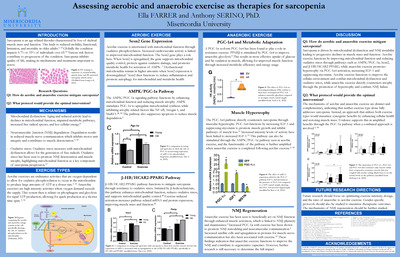
Assessing Aerobic and Anaerobic Exercise as Therapies for Sarcopenia
Ella B. Farrer
Sarcopenia is an age-related condition represented by progressive changes in muscle physiology that results in the loss of muscle mass and strength. The onset varies amongst individuals and can be influenced by various factors. On the cellular level, mitochondrial dysfunction and disruptions at neuromuscular junctions have been associated with the progression of sarcopenia. The condition ultimately reduces mobility, increases mortality, and hinders quality of life in older populations, necessitating an understanding of effective therapies.
Exercise was linked to symptom reduction of sarcopenia. Evidence indicated aerobic exercise and anaerobic exercise influence distinct molecular pathways involved in muscle physiology. Literature assessed the roles of anaerobic and aerobic exercise in influencing molecular pathways, specifically in relation to mitochondrial function, NMJ integrity, and protein synthesis.
Findings from the analysis found that aerobic exercise functions through the Sesn2, AMPK/PGC-1α, and β-HB/HCAR2-PPARG signaling pathways. Collectively, these pathways improved mitochondrial biogenesis, reduced oxidative stress, and improved cellular metabolism to limit muscle atrophy. In contrast, anaerobic exercise promoted muscle hypertrophy through PGC-1α4 pathways. PGC-1α4 was involved in the activation of IGF1, repression of myostatin, and enhanced glycolysis which increased hypertrophy. Anaerobic exercise was linked to enhanced satellite cell availability and upregulated neuromuscular communication for improved functionality.
In all, anaerobic exercise specifically targets mechanisms for muscle growth and strengthening to counteract the atrophy and muscular decline of sarcopenia, while aerobic exercise creates the cellular conditions necessary for muscular growth. Therefore, both exercise types should be utilized together to generate maximal activation of physiological pathways and most effectively mitigate sarcopenia.
-

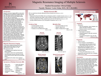
Magnetic Resonance Imaging of Multiple Sclerosis
Olivia Finlay
Magnetic resonance imaging (MRI) is a key tool in the diagnosis and monitoring of multiple sclerosis (MS). MRI allows doctors to see lesions in the brain and spinal cord and track disease activity over time. MS is an autoimmune neuroinflammatory disorder affecting the central nervous system. MS affects an estimated 900,000 people in the United States. MS most often develops in young adults, typically between the ages of 20 and 30. MS can lead to physical disability, cognitive difficulties, and a reduced quality of life. Globally, studies show that the incidence and burden of MS have increased over the past decades. This highlights the importance of early diagnosis and effective monitoring for patient outcomes. MS is treated with medication to slow the progression of the disease, steroids for flare-ups, treatment to manage symptoms, therapy to help with movement and daily activities, and healthy lifestyle support. Although MRI is effective for monitoring and detecting MS plaques, ongoing research suggests that future advancements in MRI techniques and biomarkers may improve early diagnosis and help predict disease progression more accurately.
-

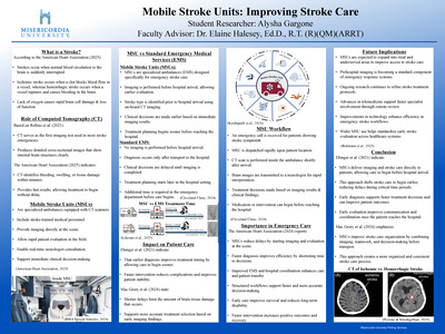
Mobile Stroke Units: Improving Stroke Care
Alysha Gargone
This project examines the role of Mobile Stroke Units (MSUs) in improving stroke diagnosis and treatment time in emergency care. Strokes occur when blood flow to the brain is interrupted, requiring rapid evaluation to reduce brain damage and improve patient outcomes. This study focuses on how MSUs, which are specialized ambulances equipped with computed tomography (CT) scanners and trained medical personnel, allow imaging and clinical decision-making to occur before hospital arrival. The project highlights the importance of CT imaging in quickly identifying stroke type, including ischemic and hemorrhagic strokes, which directly impacts treatment selection. MSU workflow allows for faster diagnosis, real-time neurologist consultation, and earlier treatment initiation compared to standard emergency medical services (EMS), where imaging and decisions are delayed until hospital arrival. Findings suggest that MSUs significantly reduce treatment delays, improve coordination between healthcare providers, and support better patient outcomes by limiting brain tissue damage. This research also identifies future implications, including expanded use of MSUs in rural areas, advancements in telemedicine, and continued improvements in emergency stroke care systems. Overall, MSUs represent an important advancement in prehospital care by shifting stroke treatment earlier in the care process.
-

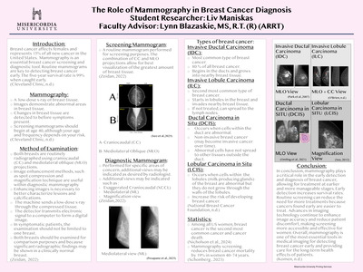
The Role of Mammography in Breast Cancer Diagnosis
Olivia J. Maniskas
Abstract Breast cancer remains one of the most common cancers affecting women, making early detection through mammography essential for improving patient outcomes. Breast cancer continues to impact millions of women each year. This capstone project shows the different types of breast cancer and their appearance on mammographic imaging. The main focus includes ductal carcinoma in situ, lobular carcinoma in situ, invasive ductal carcinoma, and invasive lobular carcinoma. The display of images demonstrate the main differences within each type of breast cancer. Each type presents with distinct imaging characteristics. This research aims to improve understanding of the various types of breast cancer and emphasize the importance of mammography for early detection and education. Increased awareness of imaging characteristics can lead to earlier detection, improved patient care, and better outcomes. This research also highlights the importance of proper positioning and image quality. The differences between screening and diagnostic mammograms are highlighted within the research and show how important taking care of your health should be.
Keywords: detection, breast cancer, increased awareness
-
 in Interventional Radiology by Silvia M. Mendoza")
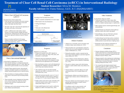
Treatment of Clear Cell Renal Cell Carcinoma (ccRCC) in Interventional Radiology
Silvia M. Mendoza
This project focuses on clear cell renal cell carcinoma (ccRCC) and the role that interventional radiology plays in treating this form of cancer. Clear cell renal cell carcinoma is the most common form of kidney cancer and is often diagnosed using diagnostic imaging such as CT scans, MRIs, and biopsies. This research includes information explaining what ccRCC is, its common symptoms, prognosis, methods of treatment, and what interventional radiology is. Some forms of treatment include surgery and immunotherapy. The main treatment of this project is minimally invasive ablation therapy. Techniques such as radiofrequency ablation, microwave ablation, and cryoablation use imaging guidance to treat tumors while limiting damage to surrounding healthy tissue. These procedures can be especially helpful for patients who may not be good candidates for surgery. Overall, this research highlights how interventional radiology is becoming an important part of cancer treatment and how minimally invasive procedures may improve a patient's outcome and survival rates.
-

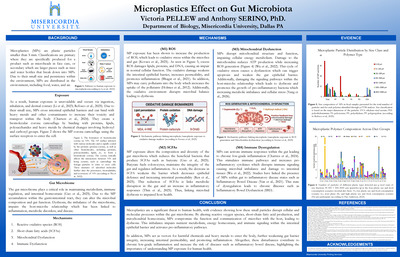
Microplastics Effect on Gut Microbiota
Victoria Rae Pellew
Microplastics Effect on Gut Microbiota
PELLEW, V, Misericordia University, 301 Lake Street, Dallas, PA, United States, 18612
Microplastics (MPs) are small plastic particles less than 5mm in size that are present throughout the environment and are commonly found in both food and water, making human exposure unavoidable. Ingestion occurs through consuming contaminated food, inhalation by breathing airborne particles, and dermal contact via phone cases or skin care products. Once absorbed into the body, MPs may influence human health by altering the gut microbiome.
Results from human fecal studies and in vitro models have shown that MPs can alter pathways associated with the reactive oxygen species (ROS), short-chain fatty acids (SCFAs), and overall mitochondrial homeostasis. Together, the alterations of the SCFAs, ROS and mitochondria dysfunction lead to inflammatory signaling, causing chronic inflammation and microbial imbalances. Over time, this exposure can negatively affect the overall health of the individual by leading to metabolic dysfunction and immune dysregulation. Current evidence highlights gut microbiota as the key mediator between MP exposure and chronic health conditions.
This paper examines current research on how MPs disrupt microbial communities, as well as how disruption within the gut contributes to immune dysregulation and metabolic dysfunction. Developing at an accelerating rate, research limitations may exist due to the limited long-term human data by means to collect fecal samples due to ethical reasoning. As a result, literature reviews highlight animal collections and in vitro systems, such as the Stimulator of the Human Intestinal Microbial Ecosystem (SHIME).
-

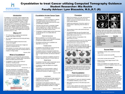
Cryoablation to Treat Cancer Utilizing Computed Tomography Guidance
Mia D. Ruckle
This research examines the effectiveness of cryoablation, a minimally invasive treatment for tumor destruction. Cryoablation works by using freezing temperatures to the targeted tissues, leading to cell damage and eventually cell death. It is being utilized more frequently to treat various cancers, such as: liver, lung, breast, renal, and prostate. This research analyzes current literature and experimental findings to evaluate how cryoablation contributes to improved treatment outcomes and reduced damage to surrounding healthy tissues. Cryoablation offers several advantages over traditional treatment options including, reduced recovery time, lower complication rates, and the ability to precisely target tumors. Imaging guidance through computed tomography during the procedure allows for accurate monitoring and control of the treatment area. The results suggest that cryoablation is an effective alternative for surgery, chemotherapy, and radiation therapy. These findings highlight the growing role of cryoablation in modern oncology. Future research should focus on long-term patient outcomes and continued advancements in imaging and procedural techniques to further improve the safety and effectiveness of cryoablation.
-

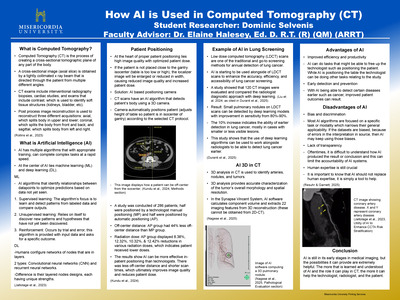
How AI Is Used in Computed Tomography
Dominic Selvenis
This project explains artificial intelligence (AI) and how it is used in computed tomography (CT). AI and CT are both defined, and the project shows how both relate to each other to create a better overall outcome of the studies. Different uses of AI are demonstrated in the following ways: patient positioning, 3D analysis, and within a lung screening study. Even though there is only one example of using AI in a lung screening study, AI can be used in almost every study to either measure, identify, or locate. Arteries, nodules, and tumors are some examples that AI can detect within CT, therefore making it an important new aspect. A few advantages of AI are in the project such as it can improve efficiency and productivity. A few disadvantages are also listed such as making sure human expertise doesn’t go away with the addition of AI. Both advantages and disadvantages give certain viewpoints to decide whether AI should stay and continue to grow in the medical imaging field.
-

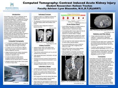
Computed Tomography: Contrast Induced Acute Kidney Injury
Keileen E. Trochez Tome
Abstract
Computed tomography is a widely used imaging modality in clinical practice due to its ability to produce detailed cross-sectional images of the body. The administration of iodinated contrast agents enhances the visualization of internal structures and diagnostic accuracy. While pre-contrast scans provide important baseline information, contrast enhanced imaging can further aid in the detection of various pathologies. However, the use of contrast media has raised concerns regarding its potential to contribute to acute kidney injury (AKI), a sudden decline in renal function typically identified in changes in creatinine levels and glomerular filtration rate (GFR). This research reviews current evidence on contrast- associated AKI. Findings from studies indicate that contrast use does not significantly increase the risk of AKI in the general population. Instead, elevated risk is primarily observed in patients with pre-existing kidney disease, reduced GFR, diabetes, advanced age, or dehydration. These results suggest that while contrast is safe, careful patient assessment remains essential. Monitoring kidney function and implementing preventative measures, such as adequate hydration, can help reduce the likelihood of renal complications in higher risk individuals. The study highlights the importance of renal complications in higher risk individuals. Future research should focus on improving risk assessment strategies and further clarifying the relationship between contrast exposure and kidney function in vulnerable population.
-

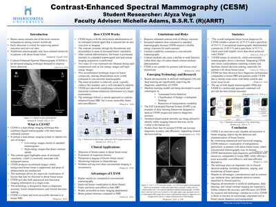
Contrast-Enhanced Spectral Mammography
Alyza Vega
Contrast-Enhanced Spectral Mammography (CESM) is an advanced breast imaging technique that enhances the detection and characterization of breast lesions, more specifically in patients with dense breast tissue where conventional mammography is limited. This research explores the principles, clinical applications, advantages, and limitations of CESM. The technique utilizes dual-energy imaging combined with intravenous iodinated contrast to highlight areas of increased vascularity, which are often seen with malignancy. CESM provides both anatomical and functional imaging in one efficient examination. Studies demonstrate that CESM offers higher sensitivity and comparable specificity to traditional mammography, with diagnostic performance approaching that of breast MRI while being more cost-effective and accessible. Clinical applications include lesion detection, preoperative staging, and monitoring treatment response. Despite its benefits, CESM does present some limitations such as increased radiation dose and risks associated with contrast administration. Emerging advancements in artificial intelligence are further enhancing diagnostic accuracy and workflow efficiency. The findings suggest that CESM is a valuable tool in breast cancer imaging and has the potential to greatly improve clinical decision-making. Future research should focus on optimizing patient selection, minimizing risks, and integrating artificial intelligence to further advance the capabilities of CESM.
-

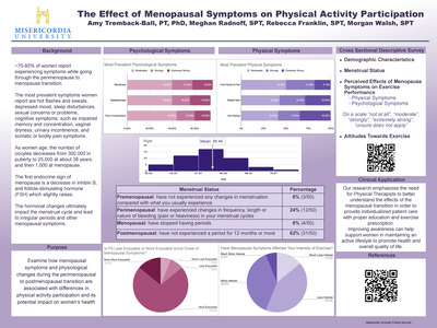
The Effect of Menopausal Symptoms on Physical Activity Participation
Morgan T. Walsh, Rebecca Franklin, Meghan E. Radnoff, and Amy Tremback-Ball